Be seen today  844-972-7752

MEDIAL BRANCH BLOCK

Do you know what a medial branch block is?

A medial branch block is a treatment we perform at Summit Spine and Joint Centers to help with pain that has developed due to a damaged or inflamed facet joint in the spine. A facet joint is the bony structure that connects spinal vertebrae together. Facet joints help control the movement of the spine and provide extra support and stability for the vertebrae.

When you have a medial branch block, a local anesthetic such as lidocaine is injected into the medial nerve of the facet joint. Injecting the local anesthetic into the medial nerve helps healthcare providers diagnose back pain. If the injection relieves at least 50% of your usual back pain, your healthcare provider will likely confirm that the pain is coming from the facet joint and recommend radiofrequency ablation as treatment. Medial branch blocks are also used to diagnose sacroiliac joint pain.

For the full article click here

If you feel pain when you are bending, twisting or flexing, you may be a candidate for a medial branch block! Call us today to begin your pain free journey! (844) 972-7752


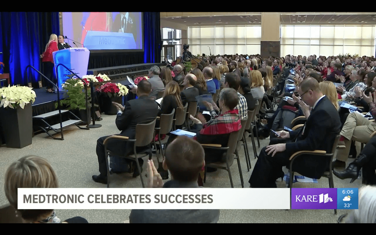
Medtronic Celebrates Successes

Medtronic Celebrates Successes I recently had the honor and privilege to present an Atlanta veteran, who is one of our patients, at the annual Medtronic holiday event as their story of the year. This patient shattered her ankle in the Army, but Georgia Pain and Wellness Center successfully placed in her a Spinal Stimulator Implant, and her pain was reduced by 90 percent. Later, in checking the serial number of the device, it turned out that it was coincidentally built in part by the veteran’s mother in Puerto Rico who works at a Medtronic factory assembling batteries for these devices. As a $125 billion company, the odds of this happening are basically an impossibility. Please feel free to share this uplifting story.
Watch Video

Why Kyphoplasty is the Best Option for Spinal Compression Fractures

Disc Herniation and Treatments Compression fractures in the spine can happen in multiple instances. It is more common than one would think. This is an injury that can effect people from all ways of life. Luckily there is a new treatment to help relieve pain and prevent the vertebrae in the spine from collapsing again. This procedure is a specialty of Dr. Patel. If you would like more information regarding how Kyphoplasty may be the right solution for your compression fractures, Click HERE.

Herniated Disc

Herniated Disc A herniated disc is a common injury that can affect any part of the spine. A herniated disc can cause severe pain and other problems in the arms or legs. A herniated disc occurs when the nucleus pushes through the outer wall. This herniation can result in a large bulge that can press against nearby nerve roots.

Herniated discs commonly result from age-related weakening of the spinal discs. This is called disc degeneration, and it can occur gradually over many years as a result of normal wear and tear on the spine. A herniated disc can also result from a traumatic injury, or from lifting a heavy object improperly.

Symptoms of a herniated disc vary depending on the location of the disc and the severity of the rupture. Some herniated discs cause no symptoms, and a person with this type of injury may not realize the disc is damaged. But a herniated disc can also cause severe pain, numbness or tingling, and weakness. Most herniated discs occur in the lower back, where they can cause symptoms in the buttocks, legs and feet.

Treatment options for herniated disc depend on the location and severity of the injury. A herniated disc may be treated with pain-relieving medications, muscle relaxers and corticosteroid injections. Visit our website for more information or give us a call at (844) 972-7752.

Chronic Back and Leg Pain, We Can Fix That.

Chronic Back and Leg Pain, We Can Fix That Whether it was a slip and fall at your job, a car wreck, or ongoing back problems we have treatment options to help chronic pain. Chronic pain can get in the way of doing normal everyday activities. Imagine if something as simple as cooking dinner for your family became too hard to do. Pain can leave you with limited options in your mobility.

One treatment option that Georgia Pain and Wellness Center offers is Spinal Cord Stimulation. This treatment option involves electrical impulses to relieve chronic pain in the back and legs. The electrical pulses prevent pain signals from being received by the brain. Spine Cord Stimulation is an option for people who suffer from neuropathic pain and for whom conservative treatments have failed.

Let one of our Board Certified Physician’s help relieve your chronic pain. Why choose us? At Georgia Pain and Wellness Center we provide compassionate care, comprehensive evaluation and testing, and proven treatment options for a variety of pain issues. Most of our testing is pain free and the minimally invasive procedures are performed in one of our four state-of-the-art surgery center. At Georgia Pain and Wellness Center, we relieve your pain and help restore your life!

What’s involved in a spine cord stimulator trial?

What is a spinal cord stimulator?

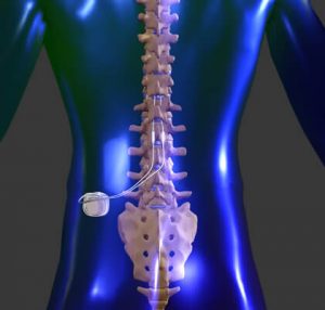
A spinal cord stimulator is a specialized device that stimulates the spinal cord and spinal nerves by tiny electrical impulses via a small electrical wire placed behind and just outside the spinal cord in the epidural space. The electrical wire or lead contains a series of four to eight evenly spaced electrodes that can be programmed to generate an electrical field.

Am I a candidate for a spinal cord stimulator?

Spinal cord stimulators are most often used for patients with chronic and severe neuropathic pain, who have not responded to other more conservative treatments. Neuropathic pain is pain due to damaged nerve tissue. Common examples include patients with post-laminectomy nerve damage, radiculopathy, diabetic neuropathy and reflex sympathetic dystrophy.

What is the purpose of a spinal cord stimulator?

Spinal cord stimulator for chronic pain.

A spinal cord stimulator generates a low level electric field to interrupt nerve conduction of pain signals to the brain.

What is the difference between a spinal cord stimulator trial and a spinal cord stimulator permanent implantation?

Typically, after patients have failed more conservative treatment options, consideration is given to a trial of spinal cord stimulation. Rather than put an expensive device permanently into a patient, a temporary spinal cord stimulator wire is placed in a patient for several days to a week. This temporary wire gives the patient an opportunity to experience spinal cord stimulation without having to undergo a full implantation. If this trial is successful in relieving pain, a permanent device can be placed under the skin at another date.

What determines if a spinal cord stimulator trial is successful?

Usually the patient who has had a trial has a very good sense of success. Most patients who are not sure that the spinal cord stimulator trial helped will not get a permanent spinal cord stimulator. Typically, the physician is looking for a significant increase in activity tolerance or a significant decrease in the need for pain medication or some combination of both.
How long does a spinal cord stimulator trial placement take?

Placement of a trial stimulator wire or lead takes from thirty minutes to an hour.

How long does a spinal cord stimulator permanent implantation take?

Placement of a permanent spinal cord stimulator wire and a subcutaneous pulse generator or battery takes from one to two hours.

How is it a spinal cord stimulator trial lead placement actually performed?

The patient is monitored with an EKG, blood pressure cuff and an oxygen-monitoring device. The procedure is performed under sterile conditions. In a spinal cord stimulator trial, temporary electrodes are placed and then the patient uses an external device to generate electrical current. The electrodes are placed under x-ray guidance with the patient lying on his belly. A local anesthetic is used to numb the skin and deeper tissues. An introducer needle is passed into the epidural space. The electrodes are inserted through the introducer needle. The position of the spinal cord stimulator electrodes is adjusted until stimulation covers as much of the painful area as possible. When this is accomplished, the introducer needle is removed and the temporary wire is secured to the skin with a small stitch and a large sticky bandage.

Will the spinal cord stimulator placement hurt?

The procedure involves inserting an introducer needle through skin and deeper tissues. There is some pain involved. Most patients also receive intravenous sedation that makes the procedure easier to tolerate.

Will I be “put out” for a spinal cord stimulator?

The placement of the trial electrodes is done under local anesthesia with patients mildly sedated. This is necessary to ensure proper placement of the wires. The amount of sedation given generally depends upon the patient tolerance. The patient is conscious although some will have enough amnesia that they may not remember parts of the procedure. In a permanent placement, once the wires or electrodes are in good position, the patient is often sedated more heavily to place the pulse generator or battery.

Where are the electrodes inserted? Where is the generator placed?

For the pain involving lower back and legs, the electrodes are inserted in the midline of the lower back, usually above any previous surgery. For a permanent stimulator, the generator is then placed on the side of the lower abdomen or above the buttock. For the pain involving the arms, the electrodes are inserted in the midline at the upper back. The generator is then placed on the side of the chest.

What should I expect after a spinal cord stimulator?

If the procedure is successful, the patient’s pain may be gone or quite less. The patient will usually feel a constant sensation of stimulation, often described as warm or tingly. The patient may have soreness due to the needle placement for a day or two. Most patients with successful stimulation take less, not more, medication after the placement.

What should I do after the procedure?

Instructions for after a spinal cord stimulator procedure.

This procedure is an outpatient procedure. The patient will need a ride home. The patient should plan to take it easy for a day or so after the procedure. The patient can do most activities but is generally advised to avoid a lot of bending or twisting of the spine.

How long will the generators last?

Depending upon the device used, the batteries may be internal or external. With internal batteries, the generator may last several years depending on the intensity and duration of stimulation required. The batteries cannot be replaced or recharged; they must be replaced surgically. With external batteries in the transmitter, the batteries can be replaced as needed.

Can a spinal cord stimulator be removed?

Yes. Even though we often consider the placement permanent, the spinal cord stimulator wire or lead can usually be removed with relative ease.

Can I have an MRI if I have a spinal cord stimulator?

No. Typically, patient with a spinal cord stimulator in place should not have MRIs. They also should not have any deep electrical or deep heating physical therapy treatments, often called diathermy. There is a risk that an MRI or diathermy may raise the temperature of the electrical wire or electrodes and potentially harm the spinal cord or spinal nerves.

Can I pass through airport security with a spinal cord stimulator?

Maybe. Depending on the sensitivity of the specific screening device, many patients can pass through with ease, just like some patients with pacemakers. If not, all patients are provided with identification indicating a medical device has been implanted.

Will the spinal cord stimulator help?

It is sometimes difficult to predict if the procedure will actually help the patient or not. For that reason, temporary electrodes are placed as a trial to determine if a permanent device will be effective to relieve pain. Typically, with successful trial stimulation, patients will have a 50 to 70% reduction in their pain.

What are the risks and side effects of spinal cord stimulation?

Generally speaking, this procedure is safe. However, with any procedure there are risks, side effects and possibility of complications. Common side effects are bruising and soreness. Less common risks are short-term weakness or numbness, headache, bleeding or infection.

Who should not have a spinal cord stimulator?

Patients on a blood thinning medication, patients with an active infection going on, and patients with poorly controlled diabetes or heart disease should not have the procedure or at least consider postponing it if postponing would improve the overall medical condition.

Call our office at (770)962-3642 to see if you may be a candidate for spine cord stimulation.

article by Dr Zeballos

Radiofrequency Ablation for Arthritis Pain

What Happens During Radiofrequency Ablation?

You will meet with a doctor for an evaluation. If radiofrequency ablation is recommended, a doctor will explain the procedure in detail, including possible complications and side effects.

The doctor will also answer any questions you may have.

An intravenous (IV) line may be placed in a vein in your arm before the procedure and a local anesthetic and mild sedative may be used to reduce any discomfort during RFA. You may be awake during the process to aid in properly assessing the procedure. Ask your doctor about specifics beforehand.

After the local anesthesia (you will be awake but will not feel any pain) has been given, the doctor will insert a small needle into the general area where you are experiencing pain. Using X-ray, your doctor will guide the needle to the exact target area. A microelectrode is then inserted through the needle to begin the stimulation process.

During the procedure, your doctor will ask if you are able to feel a tingling sensation. The object of the stimulation process is to help the doctor determine if the electrode is in the optimal area for treatment.

Once the needle and electrode placement are verified, a small radiofrequency current is sent through the electrode into the surrounding tissue, causing the tissue to heat. You should not feel discomfort during the heating portion of the procedure.

Following radiofrequency ablation:

You will stay in a recovery room for observation, where a nurse will check your blood pressure and pulse.
A bandage will be placed over the injection site.
The nurse will give you a beverage and review your discharge instructions with you.
Someone must drive you home.
Can I Resume My Normal Activities After Radiofrequency Ablation?
You will have a few restrictions immediately following radiofrequency ablation:

Do not drive or operate machinery for at least 24 hours after the procedure.
You may resume your normal diet.
Do not engage in any strenuous activity for the first 24 hours after the procedure.
Do not take a bath for one to two days after the procedure; you may shower.
You may remove any bandages in the evening before going to bed.

You may experience the following effects after RFA:

Leg numbness: If you have any leg numbness, walk only with assistance. This should only last a few hours and is due to the local anesthesia given during the procedure.
Mild back discomfort: This may occur when the local anesthetic wears off and usually lasts two or three days. Apply ice to the area the day of the procedure and moist heat the day after the procedure if the discomfort persists. You may also use your usual pain medications.

RFA Warning
If you feel severe pain at the injection site and notice swelling and redness, or increased leg weakness, have someone take you to the nearest emergency room or call 911. Tell the emergency room staff that you just had RFA. A doctor must evaluate you for bleeding and injection complications.

Lumbar Compression Fracture

Lumbar Compression Fracture The lumbar vertebrae are the 5 largest and strongest of all vertebrae in the spine. These vertebrae comprise the lower back. They begin at the start of the lumbar curve (ie, the thoracolumbar junction) and extend to the sacrum. The strongest stabilizing muscles of the spine attach to the lumbar vertebrae. Fractures of lumbar vertebrae, therefore, occur in the setting of either severe trauma or pathologic weakening of the bone. Osteoporosis is the underlying cause of many lumbar fractures, especially in postmenopausal women. Osteoporotic spinal fractures are unique in that they may occur without apparent trauma. However, a thorough diagnostic workup is always required to rule out spinal malignancy. The image below reveals a wedge compression fracture. (See Pathophysiology.)

In the past, treatment options for lumbar fractures were quite limited, with bracing and rest prescribed most often. While many patients improved with this regimen, some did not and were left with chronic, disabling pain. Suh and Lyles found that vertebral compression fractures were associated with significant performance impairments in physical, functional, and psychosocial domains in older women.[1]However, medical and surgical options are now available that can relieve the severe pain and disability from these fractures.

Pathophysiology

The lumbar spine provides both stability and support, allowing humans to walk upright. Proper function of the lumbar spine requires that it have a normal posture (ie, a normal lumbar curve). Any injury that changes the shape of a lumbar vertebra will alter the lumbar posture, increasing or decreasing the lumbar curve. As the body attempts to compensate for the alteration in the lumbar spine in order to maintain an upright posture, this will tend to distort the curves of the thoracic and cervical spine.

Lumbar compression fractures can be a devastating injury, therefore, for 2 reasons. First, the fracture itself can cause significant pain, and this pain sometimes does not resolve. Second, the fracture can alter the mechanics of the posture. Most often, the result is an increase in thoracic kyphosis, sometimes to the point that the patient cannot stand upright. In trying to maintain their ability to walk, patients with kyphosis report secondary pain in their hips, sacroiliac joints, and spinal joints. These patients are also at risk for falls and accidents, increasing the risk of secondary fractures in the spine and elsewhere.

Fractures in the lumbar spine occur for a number of reasons. In younger patients, fractures are usually due to violent trauma. Car accidents frequently cause flexion and flexion distraction injuries. Jumps or falls from heights cause burst fractures. These fractures can also result in serious neurological injury. In older patients, lumbar compression fractures usually occur in the absence of trauma, or in the context of minor trauma, such as a fall. The most common underlying reason for these fractures in geriatric patients, especially women, is osteoporosis. Other disorders that can contribute to the occurrence of compression fractures include malignancy, infections, and renal disease.

Traumatic fractures

Different types of fractures can occur in the lumbar (or thoracic) spine. Classification of these fractures is based on the 3-column anatomic theory of Denis, which describes anterior, middle, and posterior spinal columns consisting of aspects of the spine and their corresponding ligaments and other soft-tissue elements. The Denis system, however, was created to classify traumatic fractures. A similar classification system does not exist for compression fractures. The main reason to use such a classification is to help determine whether a fracture is stable. Instability in the Denis system implies that damage has occurred to at least 2 of the columns of the lumbar spine.

  • Wedge fractures are the most common type of lumbar fracture and are the typical compression fracture of malignancy or osteoporosis. They occur as a result of an axially directed central compressive force combined with an eccentric compressive force. In pure flexion-compression injuries, the middle column remains intact and acts as a hinge. Although wedge fractures are usually symmetric, 8-14% are asymmetric and are termed lateral wedge fractures.
  • Fractures involving flexion and distraction forces are often due to lap belts in motor vehicle accidents. Commonly, the posterior columns are compromised in these injuries because the ligaments of the posterior elements are disrupted. This type of injury is quite common in young children. Most patients with flexion-distraction injuries remain neurologically intact.
  • Burst fractures result from high-energy axial loads to the spine. Multiple classification systems exist for these fractures. The severity of the deformity, the severity of canal compromise, the extent of loss of vertebral body height, and the degree of neurologic deficit affect the determination of whether these injuries are unstable.

When any of the above injuries occurs with a severe rotational force, the degree of injury and of instability increases.

Nontraumatic fractures

In osteoporosis, osteoclastic activity exceeds osteoblastic activity, resulting in a generalized decrease in bone density. The osteoporosis weakens the bone to the point that even a minor fall on the tailbone, causing an axial load or flexion, results in one or more compression fractures. The fracture is usually wedge shaped. Without correction, a wedge fracture invariably increases the degree of kyphosis.

Malignancies that result in spinal fractures are most commonly metastases rather than primary bone cancers. Primary cancers that often spread to the spine via hematologic dissemination include cancers of the prostate, kidneys, breasts, and lungs. Melanoma is a less common but more aggressive cause of spinal metastasis. The most common primary cancer of the spine is multiple myeloma, but others, including a variety of sarcomas,[2] can also manifest as a spinal fracture. Nonmalignant lesions that can cause fractures include aneurysmal bone cyst and hemangioma.

Spinal infections usually start in the lumbar intervertebral disk. From the disk, the infection spreads to bone, resulting in osteomyelitis. Severe pain is the hallmark symptom. The exception is spinal tuberculosis or Pott disease. In this case, the disk spaces are typically spared and a compression fracture may be the initial manifestation that leads to its discovery.

Lower Back Pain Symptoms and Causes

Lower Back Pain Symptoms and Causes

Lower back pain can be caused by a variety of problems with any parts of the complex, interconnected network of spinal muscles, nerves, bones, discs or tendons in the lumbar spine. Typical sources of low back pain include:

  • The large nerve roots in the low back that go to the legs may be irritated
  • The smaller nerves that supply the low back may be irritated
  • The large paired lower back muscles (erector spinae) may be strained
  • The bones, ligaments or joints may be damaged
  • An intervertebral disc may be degenerating

An irritation or problem with any of these structures can cause lower back pain and/or pain that radiates or is referred to other parts of the body. Many lower back problems also cause back muscle spasms, which don’t sound like much but can cause severe pain and disability.

While lower back pain is extremely common, the symptoms and severity of lower back pain vary greatly. A simple lower back muscle strain might be excruciating enough to necessitate an emergency room visit, while a degenerating disc might cause only mild, intermittent discomfort.

Identifying the symptoms and getting a diagnosis that pinpoints the underlying cause of the pain is the first step in obtaining effective pain relief.

Common Lower Back Pain Causes in Adults

Certain causes of lower back pain have a tendency to occur more often in younger individuals versus older adults:

  • Younger adults (30 to 60 year olds) are more likely to experience back pain from the disc space itself (e.g. lumbar disc herniation or degenerative disc disease) or from a back muscle strain or other soft tissue strain.
  • Older adults (over 60) are more likely to suffer from pain related to joint degeneration (such as osteoarthritis or spinal stenosis) or from a compression fracture.

When Back Pain May Be a Medical Emergency
In cases where immediate treatment is a required, physicians will investigate possible serious causes of the pain, including any type of spinal infection, tumor or fracture.

Most cases of low back pain do not require urgent care, but anyone should see a doctor immediately if low back pain is a result of trauma, or if pain is accompanied by any of the following symptoms:

  • Fever and chills
  • Unexplained recent weight loss
  • Significant leg weakness
  • Sudden bowel and/or bladder incontinence—either difficulty passing urine or having a bowel movement, or loss of control of urination or bowel movement (cauda equina syndrome)
  • Severe, continuous abdominal pain (abdominal aortic aneurysm)

In cases where immediate treatment is a required, physicians will investigate possible serious causes of the pain, including any type of spinal infection, tumor or fracture.

Typically, younger individuals (30 to 60 year olds) are more likely to experience back pain from a lower back muscle strain or from within the disc space itself – such as a lumbar disc herniation or lumbar degenerative disc disease.

The physicians at Georgia Pain and Wellness Center are Board Certified and specialize in diagnosing and treating your back pain. Call us for back pain relief today! (770)962-3642.

Pain Medication Health Center (1) – About Celebrex (Celecoxib) COX-2 Inhibitor

Since the September 2004 recall of Vioxx, recent studies have raised questions about the potential side effects of COX-2 inhibitors. For more information, please see Understanding COX-2 Inhibitor Side Effects.

Celebrex is the brand name of a prescription pain medication in the COX-2 inhibitor class of drugs. The active ingredient in Celebrex is celecoxib. Celebrex works by reducing inflammation, which often is a component of arthritis, back pain and neck pain.

Brand name

Celebrex

Active ingredient

Celecoxib

Category

NSAID (non-steroidal anti-inflammatory drug)

Company

Pfizer Inc.

FDA approval

December 31, 1998

Indications for Celebrex

Celebrex is approved use for osteoarthritis, rheumatoid arthritis in adults, painful menstrual cramps, and acute pain in adults.

Contraindications for Celebrex

Patients who should not take Celebrex include:

  • Patients allergic to celecoxib or any other ingredients of Celebrex
  • Patients allergic to sulfonamide-type drugs or sulfa
  • Persons who have experienced asthma, hives, or allergic reactions after taking aspirins or other NSAIDs
  • Women who are in late pregnancy

Article continues below

Dosage of Celebrex

Celebrex may be taken as a 100 mg or 200 mg pill. Dosage of Celebrex is based on the patient’s medical condition and response to therapy.

The FDA has recommended the following dosages for Celebrex:

  • For osteoarthritis, oral dosage of 100 mg twice daily or 200 mg once daily.
  • For rheumatoid arthritis, oral dosage of 100 mg twice daily to 200 mg twice daily.
  • For acute pain or painful menstrual cramps, oral dosage of 400 mg initially followed by 200 mg the same day if needed. Oral dosage of 200 mg twice daily thereafter.

Patients should always be careful to not take more than the prescribed dosage of the medication.

Usage of Celebrex

Celebrex may be taken with or without food.

If a dose of Celebrex is missed, the patient may take it as soon as remembered, unless it is near the time of the next dose, in which case the patient should skip the missed pill and take the next dose at the right time.

Celebrex should be stored at room temperature and away from light and moisture.

Possible Side Effects and Warnings for Celebrex

As with all NSAIDs, there is a risk of stomach problems when taking Celebrex. Although this is a very rare side effect with Celebrex, it is still possible, and the risk increases with prolonged use.

Celebrex and other NSAIDs may cause stomach ulcers that bleed,1 and patients should stop using Celebrex and call their doctor if they experience any of the following symptoms:

  • Burning pain in the stomach
  • Dark stools that look like tar or blood in the stools
  • Vomit resembling blood or coffee grounds

Celebrex and other NSAIDs can also lead to liver damage. Warning signs include nausea, vomiting, tiredness, appetite loss, itching, yellow coloring of skin or eyes, flu-like symptoms and dark urine. Patients experiencing any of these symptoms should discontinue use of Celebrex immediately and consult their physician.1

Recent study results for Celebrex are inconclusive, but some studies show an increased risk for cardiovascular problems. More information is available at Vioxx Recall and NSAID Side Effects.

In addition to liver damage, stomach problems and heart problems, other rare but serious side effects of Celebrex include serious allergic reactions and kidney problems.

Some less serious side effects of Celebrex can include headache, indigestion, upper respiratory tract infections, diarrhea, sinus inflammation, stomach pain, and nausea.

The most common Celebrex side effects experienced by patients participating in clinical studies included stomachache, diarrhea, and headache.

General precautions

In light of new concerns regarding potential NSAID side effects, patients currently taking Celebrex should talk to their physicians to evaluate their individual treatment needs and risk factors (such as cardiovascular problems). Additional patient information can be found at Safe Use of COX-2 Inhibitors and Other NSAIDs.

Patients should never take Celebrex or any other medications without first discussing it with their doctor. It is important to discuss any other medications that are being taken (including those taken without a prescription and vitamins/supplements), and to be complete and accurate in providing a medical history to the doctor.

Patients who have any reaction to aspirin (such as asthma) or other NSAIDs (such as ibuprofen or naproxen) may be sensitive to Celebrex and other COX-2 inhibitors.

Daily use of alcohol or excessive alcohol consumption may increase the patient’s risk of serious stomach problems.

Extra caution is warranted for elderly patients taking Celebrex, as elderly patients tend to be more susceptible to possible side effects.

It is not yet known whether or not Celebrex can pass through breast milk and affect infants who are breastfeeding.

Important update on Celebrex and other NSAIDs

Since the original publication of this article, the potential side effects of COX-2 inhibitors and other NSAIDs have received heightened attention from the FDA and the drugs’ manufacturers. The FDA has issued new guidelines for prescribing this class of drugs and has called for additional research.

Treatment Option for Neuropathic Pain (4)

SPINAL CORD STIMULATORS AND PAIN PUMPS – IMPLANTABLE SYSTEMS FOR NEUROPATHY

When oral medications and/or nerve blocks do not sufficiently control the pain, advanced pain therapies or implantable systems may be effective for treating neuropathy. These systems are designed to interrupt transmission of pain signals from the spinal cord to the brain. If the neuropathic pain signals do not reach the brain, then the patient does not actually feel the pain.

  • Spinal cord stimulation (SCS) for pain control introduces low levels of electrical current to the dorsal portion of the spinal cord to block the sensation of pain. The device is implanted during a surgical procedure, and may include a fully implanted system or a system with an external power source. Spinal cord stimulators may be used to manage pain from failed back surgery syndrome or radiculopathy (sciatica or leg pain). Current medical literature indicates that 50 to 60 percent of patients will get a 50 percent or better pain relief with this procedure that has lasted up to 20 years post implant.
  • Spinal pumps (or pain pump) delivers pain medication (typically morphine) directly to the intrathecal space around the spinal cord via an implanted pump. The pump is implanted during a surgical procedure, and medication in the pump is added periodically (e.g. monthly) by injecting medication through the skin into the pump reservoir.Spinal pumps may be used to manage chronic pain from osteoporosis or axial somatic pain (nociceptive pain), and sometimes may by used to manage pain from failed back surgery syndrome (although the efficacy for this use is unclear). Spinal pumps are also used to treat painful spasticity as is seen in multiple sclerosis.

    Often multiple medications are put into the pump to treat certain specific situations. That is, morphine to treat the nociceptive pain and local anesthetics (such as bipuvicaine) to treat a neuropathic pain component.

For each of the above procedures, a trial is first performed to see if it is effective and how the patient reacts before the surgery is performed. Both of the procedures are reversible and the implantable system can be removed.

What is Radiofrequency neurotomy

What is Radiofrequency neurotomy

Radiofrequency neurotomy is a procedure to reduce back and neck pain. Heat generated by radio waves is used to target specific nerves and temporarily interfere with their ability to transmit pain signals.

The radio waves are delivered to the targeted nerves via needles inserted through the skin above your spine. Imaging scans are used during radiofrequency neurotomy to help the doctor position the needles precisely.

Radiofrequency neurotomy works better in some people than in others. Tests may be needed to determine if the nerves commonly targeted by radiofrequency neurotomy are the same nerves responsible for your pain.

Radiofrequency neurotomy is usually done by doctors who specialize in treating pain. The goal is to temporarily reduce chronic pain in the back or neck that hasn’t been adequately relieved by other means, such as medications or physical therapy.

Radiofrequency neurotomy might be recommended to treat neck pain associated with whiplash. The procedure also might be recommended for back pain that:

  • Occurs on one or both sides of your lower back
  • Extends into the buttock region and thighs but not usually past the knee
  • Worsens if you twist or lift something
  • Improves when you’re lying down

Common side effects include:

  • Temporary numbness
  • Pain at the procedure site

Rarely, more-serious side effects occur, including:

  • Bleeding
  • Infection
  • Long-term numbness
  • Paralysis
  • Nerve injury

To determine if you’re a good candidate for radiofrequency neurotomy, your doctor may perform a test to see if the nerves commonly targeted by the procedure are the same nerves responsible for your pain.

This test involves injecting a small amount of numbing medication into the precise spots where the radiofrequency needles will go. If your pain significantly lessens, radiofrequency treatment at those spots may help you.

Before the procedure, you should:

  • Avoid chewing gum or using tobacco products on the day of the procedure
  • Avoid using deodorants, lotions or perfumes on the day of the procedure
  • Use clear liquids to take any medications up to two hours before your procedure
  • Arrange for someone to drive you home

During radiofrequency neurotomy

Radiofrequency neurotomy is an outpatient procedure, so you’ll go home later that same day. The procedure usually takes about an hour.

You’ll wear a hospital gown and lie on your stomach on an X-ray table. A special X-ray machine (fluoroscope) will help the doctor position the radiofrequency needles precisely — so only the targeted nerve tissue will be affected.

An intravenous (IV) line will be placed in your arm or hand to deliver medication that will keep you comfortable during the procedure. Numbing medication will be injected into your skin before the radiofrequency needles are inserted.

After radiofrequency neurotomy

You’ll be taken to another room to rest until you feel ready to go home. You might notice some soreness in the area where the needles were inserted, but this usually goes away in a day or two.

When you get home:

  • Use an ice pack on the injections sites if you have discomfort. Your doctor may recommend using the ice pack for 20 minutes three or four times during the first 24 hours after your procedure.
  • Avoid using a heating pad on the injection sites.
  • Avoid baths for two days. You may take a warm shower 24 hours after your procedure.

Radiofrequency works better for some people than others. A key factor is whether the nerves targeted by the procedure are the same nerves responsible for your pain.

Symptom relief is temporary, usually lasting six to nine months. Repeated treatments generally have similar results.


REQUEST YOUR NEW PATIENT APPOINTMENT