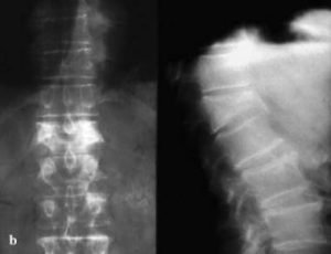
In the past, treatment options for lumbar fractures were quite limited, with bracing and rest prescribed most often. While many patients improved with this regimen, some did not and were left with chronic, disabling pain. Suh and Lyles found that vertebral compression fractures were associated with significant performance impairments in physical, functional, and psychosocial domains in older women.[1]However, medical and surgical options are now available that can relieve the severe pain and disability from these fractures.
Pathophysiology
The lumbar spine provides both stability and support, allowing humans to walk upright. Proper function of the lumbar spine requires that it have a normal posture (ie, a normal lumbar curve). Any injury that changes the shape of a lumbar vertebra will alter the lumbar posture, increasing or decreasing the lumbar curve. As the body attempts to compensate for the alteration in the lumbar spine in order to maintain an upright posture, this will tend to distort the curves of the thoracic and cervical spine.
Lumbar compression fractures can be a devastating injury, therefore, for 2 reasons. First, the fracture itself can cause significant pain, and this pain sometimes does not resolve. Second, the fracture can alter the mechanics of the posture. Most often, the result is an increase in thoracic kyphosis, sometimes to the point that the patient cannot stand upright. In trying to maintain their ability to walk, patients with kyphosis report secondary pain in their hips, sacroiliac joints, and spinal joints. These patients are also at risk for falls and accidents, increasing the risk of secondary fractures in the spine and elsewhere.
Fractures in the lumbar spine occur for a number of reasons. In younger patients, fractures are usually due to violent trauma. Car accidents frequently cause flexion and flexion distraction injuries. Jumps or falls from heights cause burst fractures. These fractures can also result in serious neurological injury. In older patients, lumbar compression fractures usually occur in the absence of trauma, or in the context of minor trauma, such as a fall. The most common underlying reason for these fractures in geriatric patients, especially women, is osteoporosis. Other disorders that can contribute to the occurrence of compression fractures include malignancy, infections, and renal disease.
Traumatic fractures
Different types of fractures can occur in the lumbar (or thoracic) spine. Classification of these fractures is based on the 3-column anatomic theory of Denis, which describes anterior, middle, and posterior spinal columns consisting of aspects of the spine and their corresponding ligaments and other soft-tissue elements. The Denis system, however, was created to classify traumatic fractures. A similar classification system does not exist for compression fractures. The main reason to use such a classification is to help determine whether a fracture is stable. Instability in the Denis system implies that damage has occurred to at least 2 of the columns of the lumbar spine.
- Wedge fractures are the most common type of lumbar fracture and are the typical compression fracture of malignancy or osteoporosis. They occur as a result of an axially directed central compressive force combined with an eccentric compressive force. In pure flexion-compression injuries, the middle column remains intact and acts as a hinge. Although wedge fractures are usually symmetric, 8-14% are asymmetric and are termed lateral wedge fractures.
- Fractures involving flexion and distraction forces are often due to lap belts in motor vehicle accidents. Commonly, the posterior columns are compromised in these injuries because the ligaments of the posterior elements are disrupted. This type of injury is quite common in young children. Most patients with flexion-distraction injuries remain neurologically intact.
- Burst fractures result from high-energy axial loads to the spine. Multiple classification systems exist for these fractures. The severity of the deformity, the severity of canal compromise, the extent of loss of vertebral body height, and the degree of neurologic deficit affect the determination of whether these injuries are unstable.
When any of the above injuries occurs with a severe rotational force, the degree of injury and of instability increases.
Nontraumatic fractures
In osteoporosis, osteoclastic activity exceeds osteoblastic activity, resulting in a generalized decrease in bone density. The osteoporosis weakens the bone to the point that even a minor fall on the tailbone, causing an axial load or flexion, results in one or more compression fractures. The fracture is usually wedge shaped. Without correction, a wedge fracture invariably increases the degree of kyphosis.
Malignancies that result in spinal fractures are most commonly metastases rather than primary bone cancers. Primary cancers that often spread to the spine via hematologic dissemination include cancers of the prostate, kidneys, breasts, and lungs. Melanoma is a less common but more aggressive cause of spinal metastasis. The most common primary cancer of the spine is multiple myeloma, but others, including a variety of sarcomas,[2] can also manifest as a spinal fracture. Nonmalignant lesions that can cause fractures include aneurysmal bone cyst and hemangioma.
Spinal infections usually start in the lumbar intervertebral disk. From the disk, the infection spreads to bone, resulting in osteomyelitis. Severe pain is the hallmark symptom. The exception is spinal tuberculosis or Pott disease. In this case, the disk spaces are typically spared and a compression fracture may be the initial manifestation that leads to its discovery.
